
Scanning Probe Microscopy (SPM) (Funded by DST-PURSE)
Instrument Details
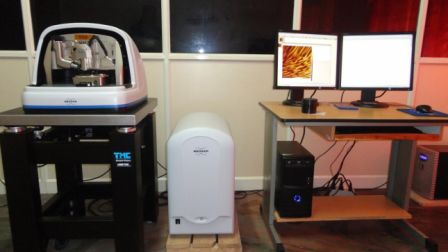
Make : Bruker Dimension Edge SPM
Application
(i) Cell studies (cancer, infectious disease). (ii) Distinguish cancer cells and normal cells based on a hardness of cells. (iii) To evaluate interactions between a specific cell and its neighboring cells in a competitive culture system. (iv) Protein imaging and crystallization, Protein/peptide interaction (v) Virus detection, Bacterial imaging. (vi) Studying dissolution rates of crystalline drugs. (vii) Identifying drug-excipient interactions. (viii) To determine encapsulation efficiency of liposomes
Advantages
(i) AFM is comparable to SEM for the fact that both are used to analyse surface topography, but AFM is superior to SEM as a 3D image of surface is possible with SPM.(ii) SPM can be used on light-atom (biological or organic) samples without special preparation, which is not possible with SEM (they need conductive surfaces – metallic or at least metal or semi-metal compounds such as oxides). (iii) AFM can even be used on surfaces under liquid. The resolution limit therefore is solely defined by the sharpness of the tip, that can be in the range of single atoms. Furthermore, the tip can be modified or functionalized with special molecules to really measure different inter-molecular or inter-atomic interactions. (Only dry samples are accepted at our facility).
Fee details and submission of application
For fee details, please visit https://kusicc.ac.in/analysis-rates/. The request form for the instrument shall be downloaded from https://kusicc.ac.in/downloads/. Read the instructions given in the form and at the bottom of the analysis rates page carefully before submitting the application. Proper method and area of focus for result has to be mentioned for accuracy.
Location: Central Laboratory for instrumentation and facilitation, University of Kerala, Karivattom campus Trivandrum